
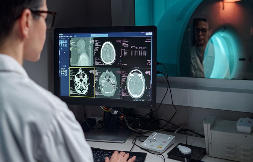
Modern healthcare is built on a simple but essential principle: successful treatment depends on an accurate diagnosis. In the past, doctors often relied on physical examinations, reported symptoms, and, in complex cases, exploratory surgery to determine what was happening inside the body. In the opinion of Kasey McKillip, the development of advanced diagnostic imaging has transformed this approach. Technologies like Magnetic Resonance Imaging (MRI), Computed Tomography (CT), and ultrasound provide detailed views of internal structures and biological activity, helping healthcare professionals make more informed and timely decisions.
Kasey McKillip offers a general overview on how diagnostic imaging supports better medical decisions
Diagnostic imaging is known for its ability to identify abnormalities before noticeable symptoms develop. Screening methods like mammography and low-dose CT scans can reveal early signs of cancer, including small lesions, nodules, and micro-calcification. Finding a disease at an earlier stage can give doctors more opportunities to manage it effectively. Early diagnosis may also reduce the need for extensive treatment, limit complications, and improve long-term outcomes. Instead of waiting for symptoms to become severe, physicians can use imaging findings to take preventive or proactive action.
Read More: NDIS Plan Review: How to Prepare and What to Request
Many medical conditions produce similar symptoms, making diagnosis challenging. For example, chest pain may result from a muscle injury, a pulmonary embolism, or a heart attack. Diagnostic imaging helps physicians distinguish between these possibilities by providing detailed structural and functional information.
As per Kasey McKillip, MRI is particularly valuable for examining soft tissues and can help identify neurological abnormalities, ligament injuries, and joint problems. Multi-slice CT scans produce rapid cross-sectional images, making them especially useful in emergencies where internal bleeding or organ damage must be identified quickly. Ultrasound, meanwhile, uses sound waves to examine tissues, organs, and blood flow without exposing patients to ionizing radiation.
Every patient has a unique anatomy and medical condition. Advanced imaging allows healthcare teams to design treatment strategies around individual needs rather than relying solely on generalized approaches. For example, three-dimensional imaging can help surgeons understand the size and location of a tumor and its relationship with nearby organs and blood vessels. This information can support more precise surgical planning. Image-guided procedures can also assist specialists during biopsies, tumor ablations, and stent placements, often reducing the need for invasive open surgery and supporting faster recovery.
Imaging remains valuable even after an initial diagnosis. Repeated scans allow doctors to determine whether a treatment is producing the expected results. In cancer care, PET and CT scans can help track changes in tumor activity and size during treatment. If a therapy is proving ineffective, clinicians can make timely adjustments. Similarly, MRI can help monitor chronic conditions affecting the brain, joints, and heart. Regular imaging provides physicians with information about disease progression and can support earlier intervention when new changes appear.
Read More: Tennessee Men’s Clinic Discusses Fitness Habits That Support Hormone Optimization
Diagnostic imaging can also strengthen communication between healthcare professionals and patients. When patients are shown images of a fracture, tumor, ligament tear, or other abnormality, complex medical explanations often become easier to understand. Visual evidence can reduce confusion, improve confidence in the diagnosis, and encourage patients to follow recommended treatment plans.








+ There are no comments
Add yours